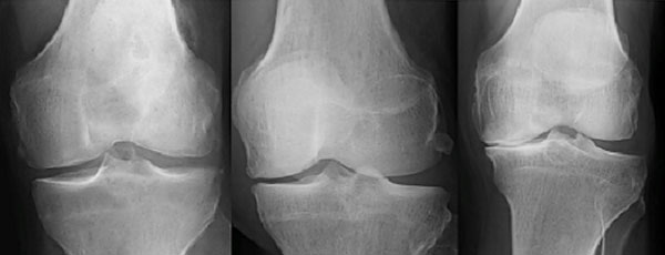
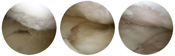
These three x-rays (above) demonstrate along with the arthroscopy (key-hole surgery) photographs (below) that arthritis can present in different ways for different patients. Moving from left to right shows increasing levels of severity of arthritis (mild, moderate, severe).
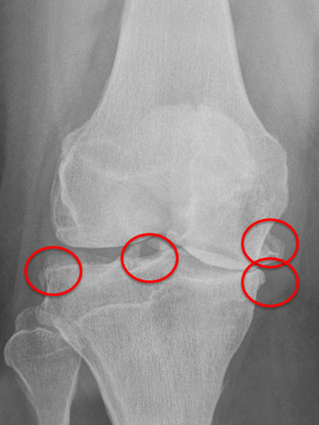
The x-ray below has been taken looking at the knee from front to back. It shows bone on bone contact on the inside of the joint. It also demonstrates osteophytes. Osteophytes are formed to try and repair the joint and take load away from the damaged area. They are normally found at the edges or periphery of the joint. The osteophytes have been highlighted.
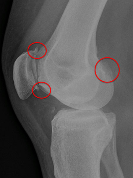
The next x-ray of the same patient but taken from the side of the knee shows different osteophytes to the ones seen in the previous x-ray. This x-ray also shows that the kneecap joint is affected with arthritis as well.
The last 2 x-rays shows the end stage of arthritis when the joint itself starts to collapse and bone is contacting bone. This patient’s joint was very stiff and sore to move. This patient has then gone onto total knee joint replacement.

