What is it?
A multi-ligament knee injury (MLKI) occurs when two or more ligaments around the knee are injured. It is also referred to as a knee dislocation. It normally occurs from a high-energy force or trauma in young people but in older people the force does not need to be as great. Any two ligaments may be involved.
What ligaments are involved?
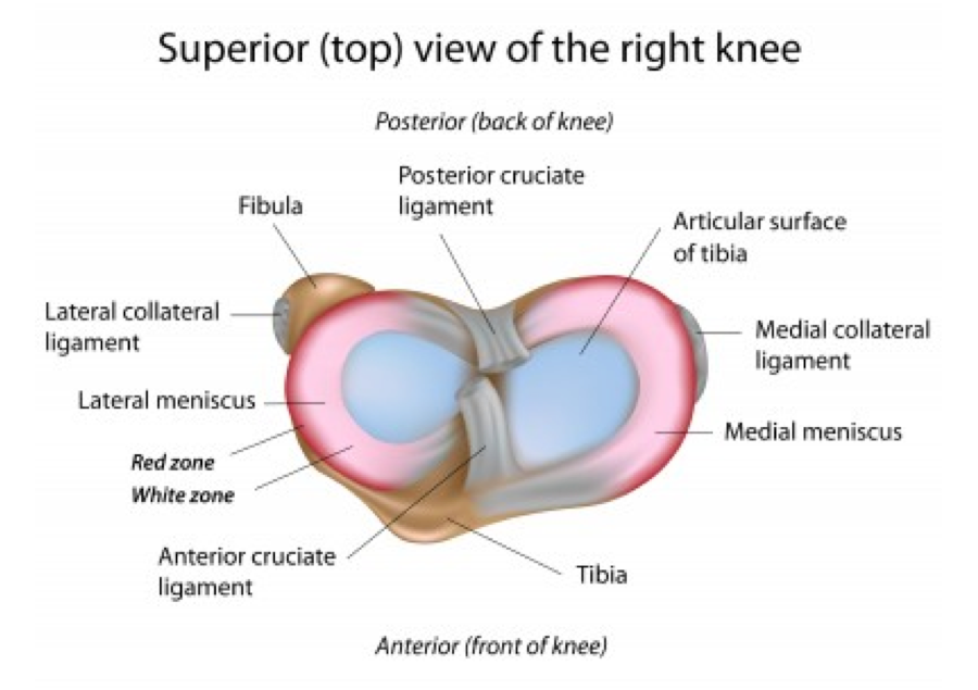
Ligaments are soft tissue structures that connect one bone to another bone on either side of a joint. They help the joint with stability and control movement. In the knee there are:
Two cruciate ligaments
- Anterior Cruciate Ligament (ACL)
- Posterior Cruciate Ligament (PCL)
Two collateral ligaments
- Medial Collateral Ligament (MCL)
- Lateral Collateral Ligament (LCL)
Two posterior ligament complexes
- Postero Lateral Corner (PLC)
- Postero Medial Corner (PMC)
Generally speaking for a knee dislocation or MLKI to occur you have to rupture either one or both cruciate ligaments along with one or both collateral ligaments.
Depending on the type of injury pattern you may also rupture one of the posterior ligament complexes.
How does it occur?
Generally from high energy forces or velocities and are normally the result of trauma:
- Sports
- Motor vehicle accident
- Workplace accident
In the older or obese population the forces needed to rupture ligaments around the knee are not as great so may occur from everyday events. Thankfully though MLKI is a rare event.
What happens?
- In a knee dislocation or MLKI, the knee literally dislocates.
- Most knees reduce spontaneously however there are some that require emergent reduction either in the emergency department or an operating theatre.
- There is obviously immediate pain and a large swelling will develop.
- The pain is far beyond what occurs with any other injury to the knee.
- There may be an obvious deformity.
- The patient will say that their knee felt as though bones had moved, that it felt like it dislocated, or went in a strange direction.
- You may have some tingling, numbness, or weakness below the knee as stretching or rupturing of a nerve can occur.
- In a small percentage of knee dislocations there may be damage to the artery at the back of the knee. This may result in the leg and foot of the patient going cold and/or changing colour due to the restricted blood flow.
- The patient will present to the emergency department and will be assessed thoroughly. Sometimes however a diagnosis is not made as the knee has already reduced and x-rays may only show very subtle signs.
Imaging
X-rays are the first choice investigation. If the knee is obviously dislocated it is wiser to reduce the knee first as this will aid the patients pain levels and then get an x-ray.
Further Imaging
If there is any chance there may be an injury to an artery the patient will have to have an arteriogram (this is where dye is injected into a blood vessel and then images are taken to see if there is any damage to the artery at the back of the knee).
Once the patient’s knee has been reduced, braced appropriately, and all other injuries excluded, then a MRI scan of the knee should be performed to assess what has occurred to the knee. Ligaments, articular cartilage (lining of the joint), menisci, tendons, and nerves will be assessed.
Surgery
Nigel will have an open and lengthy discussion with you about the implications of your MLKI. Unfortunately they do require either reconstructive surgery or repair depending on what ligaments have been damaged.