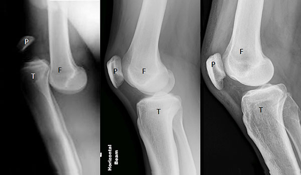
These x-rays demonstrate the range of what Doctors may see when initially seeing patients with a knee dislocation. The x-ray on the left demonstrates quite clearly an anterior knee dislocation (the tibia is sitting in front of the femur) that has not been reduced. The x-rays in the middle and on the right were also of people who had knee dislocations (but whose knees had spontaneously reduced, that is moved back into a normal position). There are several clues in the middle and right hand x-rays that demonstrate an injury to more than one ligament.
Further Imaging
If there is any chance there may be an injury to an artery the patient will have to have an arteriogram (this is where dye is injected into a blood vessel and then images are taken to see if there is any damage to the artery at the back of the knee).
Once the patient’s knee has been reduced, braced appropriately, and all other injuries excluded, then a MRI scan of the knee should be performed to assess what has occurred to the knee. Ligaments, articular cartilage (lining of the joint), meniscal cartilages, tendons, and nerves are assessed.
MRI Imaging
These MRI scans below demonstrate complete ruptures of both the Anterior and Posterior Cruciate Ligaments of the same patient. The arrows point to where both the ACL and PCL should be, but are not.
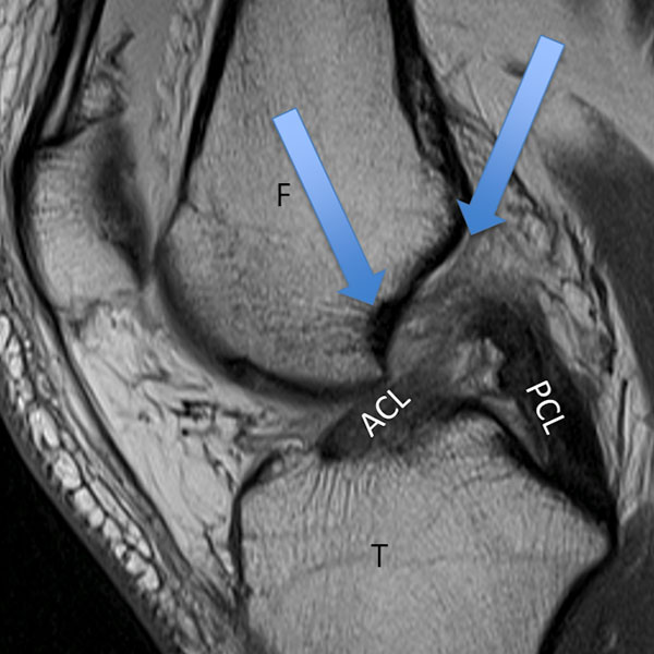
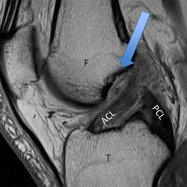
F=Femur, T=Tibia, ACL=Anterior Cruciate Ligament, PCL=Posterior Cruciate Ligament
These next MRI scans (LEFT taken from the side, RIGHT taken from the front) show a ruptured ACL and the arrow demonstrates where it should be. The red circle demonstrates a ruptured LCL and also damage to the PLC.
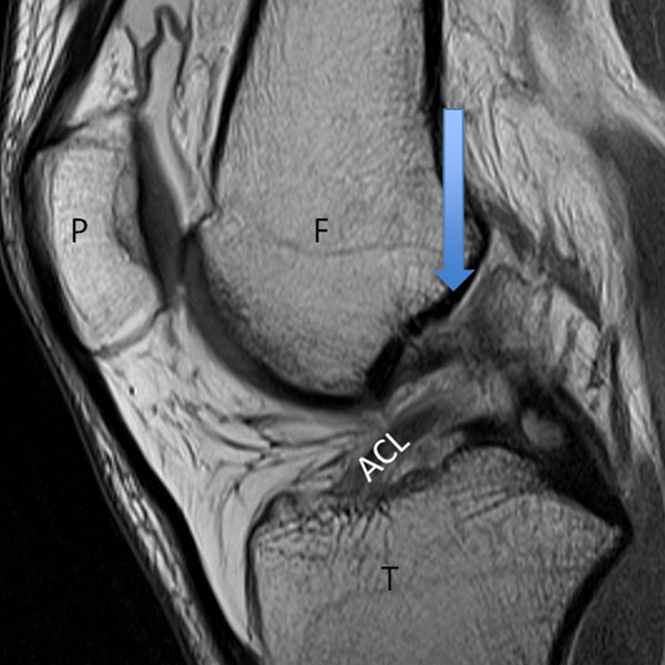
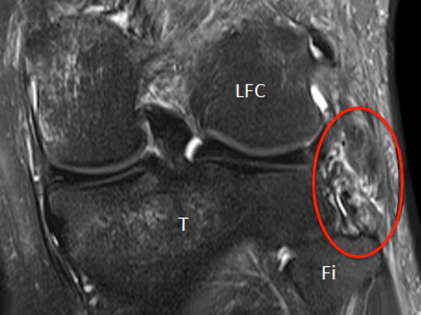
P=Patella, F=Femur, T=Tibia, ACL=Anterior Cruciate Ligament, LFC=Lateral Femoral Condyle, Fi=Fibula.